![[image]](pics/XTwave.jpg)
![[image]](pics/XTcontour.jpg)
3-D plot of pressure vs. distance along the aorta and time
contour plot of the same data
We have obtained measurements of the pressure in the human aorta in patients undergoing cardiac catheterisation. The measurements are made with dual pressure and Doppler ultrasound velocity catheter [Combowire XT (Volcano Corporation)]. After the cardiac investigation is completed 1 min of data are taken in the ascending aorta just downstream of the aortic valve. The catheter is then withdrawn 10 cm and another measurement is taken. This is repeated every 10 cm along the aorta, iliac and femoral artery down to the insertion point of the catheter.
|
|
|
|
3-D plot of pressure vs. distance along the aorta and time |
contour plot of the same data |
The data are presented as a 3-D plot of pressure vs. distance and time to the left and as a contour plot on the right. The resolution is less than the comparable data from animal experiments where the pressure was measured at 2 cm intervals, but the results are similar. The speed of propagation of the initial compression wave generated by the contraction of the left ventricle can be seen in the slope of the contours at the start of systole. The contours during diastole are much more uniform and, given their coarse resolution, show that the assumption that the diastolic fall-off in pressure is uniform throughout the arteries is not a bad one.
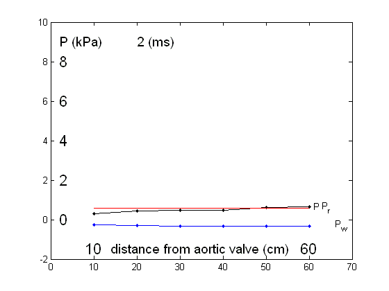
The data at each site was used to obtain the reservoir pressure with the results shown in the following figure.
![[image]](pics/ResultsAKAorta.jpg)
The top graphs show the measured pressure (left) and the calculated reservoir pressure (right) in absolute terms along 60 cm of the aorta and iliac arteries. The bottom graphs show the same data shifted relative to the diastolic (minimum) pressure and the time of the diastolic pressure. The data shown are the ensemble averages over 1 min, which includes several respiratory cycles.
The measured pressures show changes in the pressure waveform that are typical as it propagates along the large arteries. Despite these changes in wave form, the calculated reservoir pressure is remarkably uniform at all of the measurement sites.
We are most familiar with arterial pressure waveforms plotted as P(t) at different locations [example]. It is also instructive to consider P(x) at different times.
![[image]](pics/Twave_AK.jpg) |
![[image]](pics/Xwave_AK.jpg) |
|
P(t) measured at different locations of the aorta |
The same data plotted as P(x) at different times |
The figure to the right shows the measured data at 6 locations 10 cm apart along the human aorta at different times relative to the R-wave of the ECG. The solid lines start at 100 ms and the is a time interval of 10 ms between successive lines. The initial compression wavefront can be seen propagating along the aorta in the first 6 lines. Thereafter, the pressure is almost uniform along the aorta. After 360 ms the pressure waveform starts to fall and they are shown as dashed lines. The rapid fall in pressure at the end of systole is just visible in the dashed lines. During the whole of systole the pressure is extremely uniform all along the aorta.
The figure showing P(x) at different t is rather hard to interpret. It may be easier to follow the time evolution of P(x) in the movie form. These are the same data P(x) but time is shown in time; the counter shows the time in ms from the R-wave of the ECG. Also shown in the movie are the reservoir pressure (uniform along the aorta) and the wave pressure obtained by subtracting the reservoir pressure from the measured pressure.
|
|
|
|
P(t) measured at different locations of the aorta |
The same data plotted as a movie of P(x) at different times |
The propagation of the wave is seen to be very fast, taking only 50-60 ms to traverse the 50 cm between the most proximal and distal measurement sites. This is consistent with the wave speed being approximately 10 m/s in the aorta.
The previous example used ensemble-averaged data to reduce the confusion of beat-by-beat variations with variations from site to site. One of the advantages of wave intensity analysis is that it is performed in the time domain with no assumptions about the periodicity of the data. In order to illustrate this, as well as the importance of the reservoir pressure, we have applied the analysis to data obtained in the canine ascending aorta during a missing beat.
The top graph shows the ECG and simultaneously measured pressure at four sites; the ascending aorta (Asc), the aortic arch (Arch), the thoracic aorta (Thor) and the abdominal aorta (Abd); over a period of about 3 s during which there was a missing beat. The QRS complex of the ECG at 1.18 s is not accompanied by any perturbation of the arterial pressure.
![[image]](pics/missing_P.jpg) |
The measured data are shown by the thicker lines. They show the expected change in waveform as you move more distally in the aorta in the first beat which is normal. The second beat is very similar to the first, normal beat except that diastole is considerably prolonged due to the missing beat. The regularity of the exponential decay in pressure during the extended diastole is noticeable and provides further evidence against the cardiovascular system being in steady state oscillation. The third beat exhibits a very large increase in pulse pressure as would be expected from the Frank-Starling law given the increase in filling time due to the preceding missing beat. The reservoir pressure calculated from the measured pressures is shown by the thinner line. Each beat was analysed separately and the time constant of the pressure decay was very similar for all of the beats at all of the sites. Also, although it is not as evident from this view of the data, the reservoir pressures were again very similar at all of the sites along the aorta for each of the beats. |
![[image]](pics/missing_U.jpg) |
This figure shows the corresponding, simultaneously measure volume flow rates (thicker lines) during the same period together with the wave pressure (thinner lines) obtained by subtracting the calculated reservoir pressure from the measured pressure. In order to compare the different waveforms they were scaled so that their peak values were 1. In the ascending aorta, we see that there is excellent agreement between the flow and the wave pressure waveforms, particularly during systole. As we move to more distal measurement sites, the agreement remains very good during early systole but they begin to deviate more and more during late systole. We believe that this is due to the larger reflected waves seen in the more distal sites because of their proximity to the major reflection sites in the microcirculation. This is discussed in more detail in the section on wave trapping [wave trapping] |